A specific pain pattern
Intended for selected chronic low back pain associated with vertebral endplate changes—not back pain in general.
Patient Education · Spine Procedure
Basivertebral nerve ablation (BVNA, sometimes called BVN ablation) is intended for a carefully selected group of people with chronic vertebrogenic low back pain. It targets a small sensory nerve inside the vertebral body. It is different from medial branch radiofrequency ablation for some facet-joint pain.
This page is general education, not a diagnosis or a promise that a procedure is appropriate for you. Individual decisions require evaluation by a qualified clinician.
See who may be considered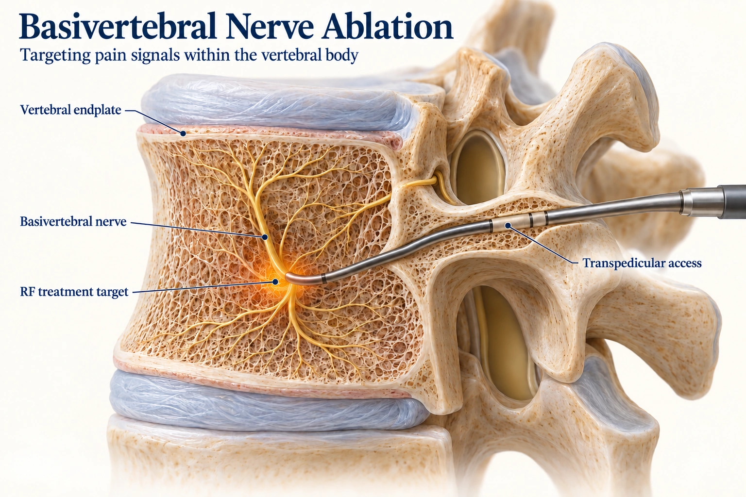
Intended for selected chronic low back pain associated with vertebral endplate changes—not back pain in general.
Modic type 1 or 2 changes may be relevant, but imaging alone cannot identify the pain source or determine candidacy.
Studies report improvements for some carefully selected patients. Relief is not guaranteed, and an individual's response cannot be predicted.
The anatomy
The basivertebral nerve travels inside the vertebral body and carries sensory information from the vertebral endplates—the thin interfaces between each vertebral body and the disc next to it. Changes around these endplates can be one contributor to chronic low back pain.
BVNA uses radiofrequency energy to ablate a targeted portion of this nerve from within the bone. It is intended to reduce pain signals from a vertebrogenic source; it does not repair the disc or treat every cause of back pain.
Patient selection
The FDA-cleared Intracept system labeling describes treatment for chronic low back pain lasting at least six months that has not responded to at least six months of conservative care, with MRI findings consistent with Modic type 1 or type 2 changes at L3 through S1. These features guide clinical evaluation; they do not prove eligibility. Other systems, clinical circumstances, and coverage policies may differ.
Feature 1
At least six months, still limiting despite a meaningful course of nonsurgical care.
Feature 2
Modic type 1 or type 2 changes at one or more vertebral levels from L3 through S1 in the Intracept labeling.
Feature 3
Symptoms, examination, and imaging are considered together while other likely pain sources are assessed.
Modic changes can be present in people without pain, and back pain can have more than one contributor. A clinician considers symptoms, examination, MRI, prior care, overall health, and alternative sources together. MRI alone cannot diagnose vertebrogenic pain.
The procedure
The care team reviews your symptoms, examination, prior treatment, and MRI or CT images. They discuss how the clinical pattern and imaging fit, alternatives, device-specific risks, medicines, and sedation. Intracept instructions call for imaging guidance and at least moderate conscious sedation; approaches may vary by system and patient.
In a sterile procedure setting, the clinician uses fluoroscopy or CT to guide specialized instruments through a small skin opening into the vertebral body. A radiofrequency probe is positioned at the target and energy is applied to ablate the basivertebral nerve. One or more levels may be treated depending on evaluation and device instructions.
The care team monitors you and gives instructions for activity, incision care, medicines, and follow-up. Temporary soreness or a short-term increase in pain can occur. Recovery and return to usual activities vary; follow your treating team’s plan rather than a general timeline.
Benefits and tradeoffs
Clinical studies of carefully selected participants report improved pain and function; follow-up studies report that some participants retained benefit at five years. Studies used specific selection criteria, and some long-term reports no longer had a randomized comparison group. Evidence cannot predict an individual result. Relief is not guaranteed, and treatment may not help if the main pain source is elsewhere.
Potential adverse events in the Intracept instructions include pain or discomfort, bleeding or hematoma, infection, nerve injury, unintended injury to nearby structures, and pedicle or vertebral-body fracture. There are also risks related to sedation or anesthesia and imaging radiation. Relevance depends on the patient, procedure, and device; discuss your individual risk with a clinician.
Options may include exercise-based rehabilitation, activity or lifestyle changes, medication when appropriate, other targeted procedures, or surgical evaluation, depending on the pain source, health, and goals. Choosing among them requires an individualized clinical discussion.
Next steps
For more on persistent back pain, see Low Back Pain Evaluation. For a different target, see medial branch radiofrequency ablation.
Device safety labeling and long-term clinical evidence inform this overview. Study results describe groups and do not establish an individual’s outcome.